Educational Training Resources offered by NoCTURN Members
CT Scanning Background
NC State University Analytical Instrument Facility: Short Introduction to X-ray Computed Tomography (Learn how CT scanners work)
Basics and Physics of X-Ray Imaging & 3D Computed Tomography (Volume Graphics (Hexagon)) (Learn why CT scanners work)
Introduction to Micro-CT Scanning introductory video for the University of Chicago PaleoCT facility. Covers basic vocabulary, parts of the machine, mounting, common artefacts, and quality/speed/resolution tradeoffs.
Background and Theory video chapter of the University of Chicago PaleoCT training course. Covers basic vocabulary, parts of the machine, common artefacts, and quality/speed/resolution tradeoffs.

Sample Preparation and Scanning
Mounting Specimens video chapter of the University of Chicago PaleoCT training course. Covers intro to mounting and wet specimens.
Pre-scan video chapter of the University of Chicago PaleoCT training course. This video covers power modes, changing a dual tube system, and warmup. This video is specific to Phoenix/GE/Waygate machines.
Scanning with a High Power 240 kV Tube video chapter of the University of Chicago PaleoCT training course. Covers scanning software for Waygate machines, positioning a specimen in the scanner, how to read a histogram, scanning parameters, and detector calibration.
Fossils and Filters video chapter of the University of Chicago PaleoCT training course. Covers tips for mounting fossils, and when and how to use filters.
Scanning with a 180 kV nano-focus tube video chapter of the University of Chicago PaleoCT training course. Covers the differences between the 240 kV micro-focus and 180 kV nano-focus tubes, mounting, positioning and parameters for lower density specimens, and additional focus tools.
Additional Scan Tips video chapter of the University of Chicago PaleoCT training course. Covers mutliscan, observation ROI, and troubleshooting.
MICRO Specimen Packaging Recommendations for Optimal Mounting (Learn how to package your samples for optimal CT imaging)

Special Sample Preparation
MICRO introduction to diceCT: Tips and tricks for diceCT staining natural history specimens, which allows for the visualization of soft-tissue structure in CT scans.
Sample Data: See what CT Data is Like
Seattle Children’s Research Institute Hosts Freely Available CT Data (from a Bruker/Skyscan 1272 micro-CT scanner —great for learning how to make 3D models in freeware tools, like 3D Slicer)
- Adult Mouse Skull (dry): 1K camera, 24 micron, scan duration 40 min. Size ~0.5GB
- Adult Mouse Skull (dry): 4K camera, 6.125 micron, scan duration 8.5h. Size ~35GB
- Iodine stained, adult mouse kidney: 1K camera, 18 micron, scan duration 40h. Size ~0.5GB
- Iodine stained, E15 whole mouse embryo: 2K, 9 micron, scan duration 1h 50m. Size ~5GB
- Full body adult mouse scan: 512 camera mode, 52.3 micron, scan duration 53 m. Size ~0.6GB
Find more data, methods, and example 3D models via the SlicerMorph literature on Google Scholar

CT Data Processing
MICRO Instructions for Working with Image Stacks in FIJI (FIJI Is Just ImageJ): Tips and tricks for manipulating CT-scanner image stacks using the free software ImageJ
3D Slicer & SlicerMorph (free, open source software for visualization, processing, segmentation, registration, and analysis of medical, biomedical, and other 3D images and meshes)
• SlicerMorph Tutorials on GitHub
• Join the 3D Slicer Discourse and be part of the conversation! (Note: Beta Testers can share feedback and feature requests with Slicer Coders.)
3D Digital Reconstruction video chapter of the University of Chicago PaleoCT training course. Covers reconstruction tools available on Datos, such as AGC, cropping, and other modules.
Data Processing Basics in VG Studio video chapter of the University of Chicago PaleoCT training course. Covers importing data, navigation, reorientation, cropping, and merging scans.
University of Florida DeLeon Lab Data Post-processing Modules (Learn how to process CT datasets for optimal use)
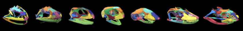
Gray’s Colors of Skull Anatomy: Do you render skulls? Stylize the color of each bone so that everyone recognizes the same element across wildly different skull shapes.
CT Data Stewardship
Research Data Alliance: Generalist Repository Comparison Chart (Learn about housing your CT data and digital models online)
Education
Collections Education Coffee Break Series: oVert TCN Resources for Education: A brief introduction to educational resources associated with the oVert project. This resource includes the slides and recording of Dr. Jaimi Gray’s presentation as well as links to all of the resources she discusses (doi:10.25334/95DJ-MY09)
3D Think Game by ERSUCC: Test your spatial reasoning with this puzzle game inspired by tomography!
- Understand what microCT data is and how to download and store this data
- Open and visualize microCT data
- Interpret microCT data
- Collect measurements from microCT data
- Generate 3D models from microCT data
[Thank you to all of our contributors! If you have training or education content that you’d like to see included here, please reach out to the Education & Outreach Committee at our Slack channel, or join the Network!]
Recent Blog Posts by the Education & Outreach Communinty
- Thousands of natural history specimens that you can access for free!
 We are in an era of digital innovation, and today the barriers to accessing the wonders of the natural world have been shattered like never before. Spearheaded by scientists at the University of Florida’s Florida Museum of Natural History, a… Read more: Thousands of natural history specimens that you can access for free!
 - By RSS, Google Alerts, in Slack, or however you like. You can now keep up with NoCTURN activitities on your terms.
 NoCTURN Blog Posts now autopopulate to the NoCTURN Slack. If you prefer, however, you can also save the URL of the whole Blog or just select categories (click the header at the top of a post for all posts in… Read more: By RSS, Google Alerts, in Slack, or however you like. You can now keep up with NoCTURN activitities on your terms.