Why are acknowledgements important? Acknowledgements are more than just a feel-good afterthought when submitting your paper: they are of material value to technical staff, shared labs, and future readers of your papers. Technical staff involved in a wide range of […]
Educational Resource / Resources
Posted on:
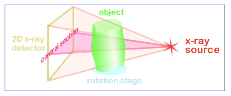
What is microCT imaging?
Click here for a GitHub resource on how micro-CT imaging works, and how it can be used to capture detailed 3D microstructural images. MicroCT imaging, short for micro-computed tomography, is a technique that uses X-rays to create detailed 3D images […]
Educational Resource / Resources
Posted on:
Practical Microcomputed Tomography Workshop
Create by Stuart Stock, hosted by the Northwestern University Feinberg School of Medicine Description This record presents the material presented at the Denver X-ray Conference (DXC), Aug 2024, in Westminster, CO. It is designed to introduce practical numerical analysis of […]
Educational Resource
Posted on:
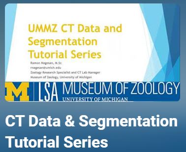
Ecology and Evolutionary Biology Museums YouTube Channel, CT Data & Segmentation Tutorial Series
By Ramon Nagesan, University of Michigan Museum of Zoology YouTube playlist of tutorials that range from introduction to CT Data, MorphoSource, and processing data in VGStudio. https://www.youtube.com/playlist?list=PLRu5Ab94NHuxquFgNafDfVY6cJPe1jWRt
Educational Resource
Posted on:
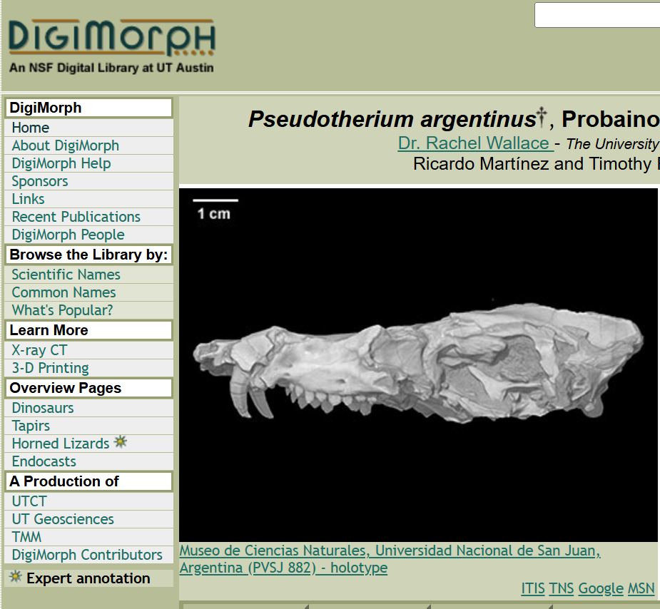
DigiMorph
By UTCT, University of Texas https://www.digimorph.org/index.phtml The Digital Morphology library is a dynamic archive of information on digital morphology and high-resolution X-ray computed tomography of biological specimens.
Educational Resource
Posted on:
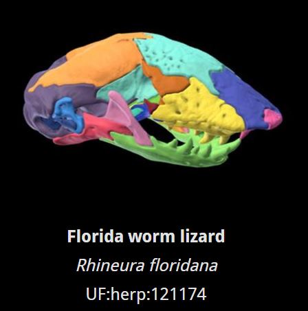
Gray’s Colors of Skull Anatomy
By Jaimi Gray, UTCT, University of Texas http://www.graysvertebrateanatomy.com/work/colorsofskullanatomy/ Standardized color scheme for rendering skulls.
Educational Resource
Posted on:
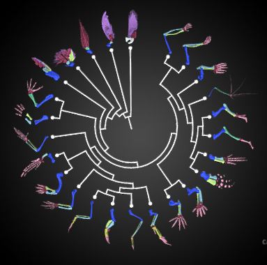
Forelimb homology 3D model collection
By Jaimi Gray, UTCT, University of Texas http://www.graysvertebrateanatomy.com/work/forelimbhomology/ A collection of colorized skeletal forelimb models from the vertebrate tree of life.
Educational Resource
Posted on:
3D Slicer Training
By 3D Slicer https://www.slicer.org/wiki/Documentation/Nightly/Training 3D Slicer’s Wiki page of tutorials.
Educational Resource
Posted on:
MICRO Specimen Packaging Recommendations for Optimal Mounting
By MICRO, University of Arkansas https://bpb-us-e1.wpmucdn.com/wordpressua.uark.edu/dist/4/453/files/2018/08/MICRO_SpecimenMounting-186j0p7.pdf Learn how to package your samples for optimal CT imaging.