
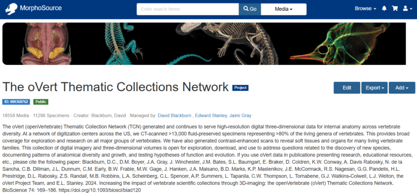
Between 2017 and 2023, the “open Vertebrate” (oVert) project, funded by NSF, united 18 participating institutions to digitize vertebrates in their natural history collections through Computed Tomography (CT) scans. Six institutions acted as imaging centers with CT scanning labs, including Harvard University, Texas A&M University, University of Chicago, University of Florida, University of Michigan, and University of Washington. Natural history collections, researchers, educators, and artists all benefit from the availability of digital 3D representations of specimens on MorphoSource. You can read more about how the oVert project formed a community and subsequently generated and shared their data in their publication in BioScience.
Data from the main oVert grant is available via MorphoSource on the oVert Thematic Collections Network project page, and additional data from Partner to Existing Networks (PEN) grants are available via their individual project pages. These include:
- FuncQEE: 1,700 species of rodents, with a focus on less common species
- GalapaGateway: Galapagos vertebrates
- oBird: Bird skins “outside of the bird” (photogrammetry)
- oMEGA: vertebrates larger then 250 kg (laser scans)
- oMeso: Mesoamerican amphibians and reptiles
- oUTCT: UTCT archive of vertebrates
- CryptoVert: small bodied cryptobenthic fishes
For each project page, you will find the media (i.e. scans) associated with oVert or one of its PENs. Near the top of the page, you will see the number of media and the number of specimens associated with the project. The number of media is higher than the number of specimens because for any given specimen, the oVert team typically generated multiple scans, including whole body scans, a higher resolution scan of the head, and for some, contrast-enhanced scans too.
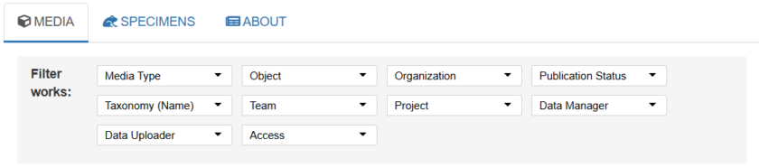
To find the scans you’re looking for, you can use the dropdown boxes at the top of the media list to filter the scans by many different fields. To view scan volumes only (i.e. exclude derivative meshes that may have been generated from scans), you can use “media type” to select “volumetric image series”. Use the other filter fields to narrow down the media list further.
Once you see something in the media list you’d like to take a look at, click on its thumbnail on the left hand side. A “media page” will open in a new tab. On the right hand side of the page, you will see a 3D viewer. You can use the “tools” menu on the left hand side of the 3D viewer to view the contents of the scan (switch the “mode” to “volume” to see a volume rendering and adjust the windowing sliders until you see the contents). You will see a low resolution version of the scan, and you can use this to confirm it contains your structures of interest.
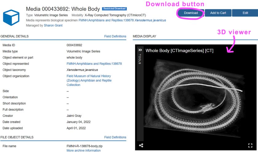
All oVert scans are available for download (with more still being uploaded!) and are listed under two categories. On the project page, in the “publication status” column on the right hand side, you will see scans listed as either:
Open download: when you click the download button, you will need to fill out some fields prior to download. You will then be able to download the data immediately.
or
Restricted download: when you click the download button, you will need to provide some information about how you will use the data. A download request will be sent to the manager of the data. Once they approve it, it will be available to download in your MorphoSource dashboard under “requests for media” in the “my downloads” section.
Their publication status depends on the institution that specimen came from, as it is up to the collections staff to make decisions about how to best manage their data. Typically all download requests will be approved, as long as you’re not a bot, or up to anything nefarious!