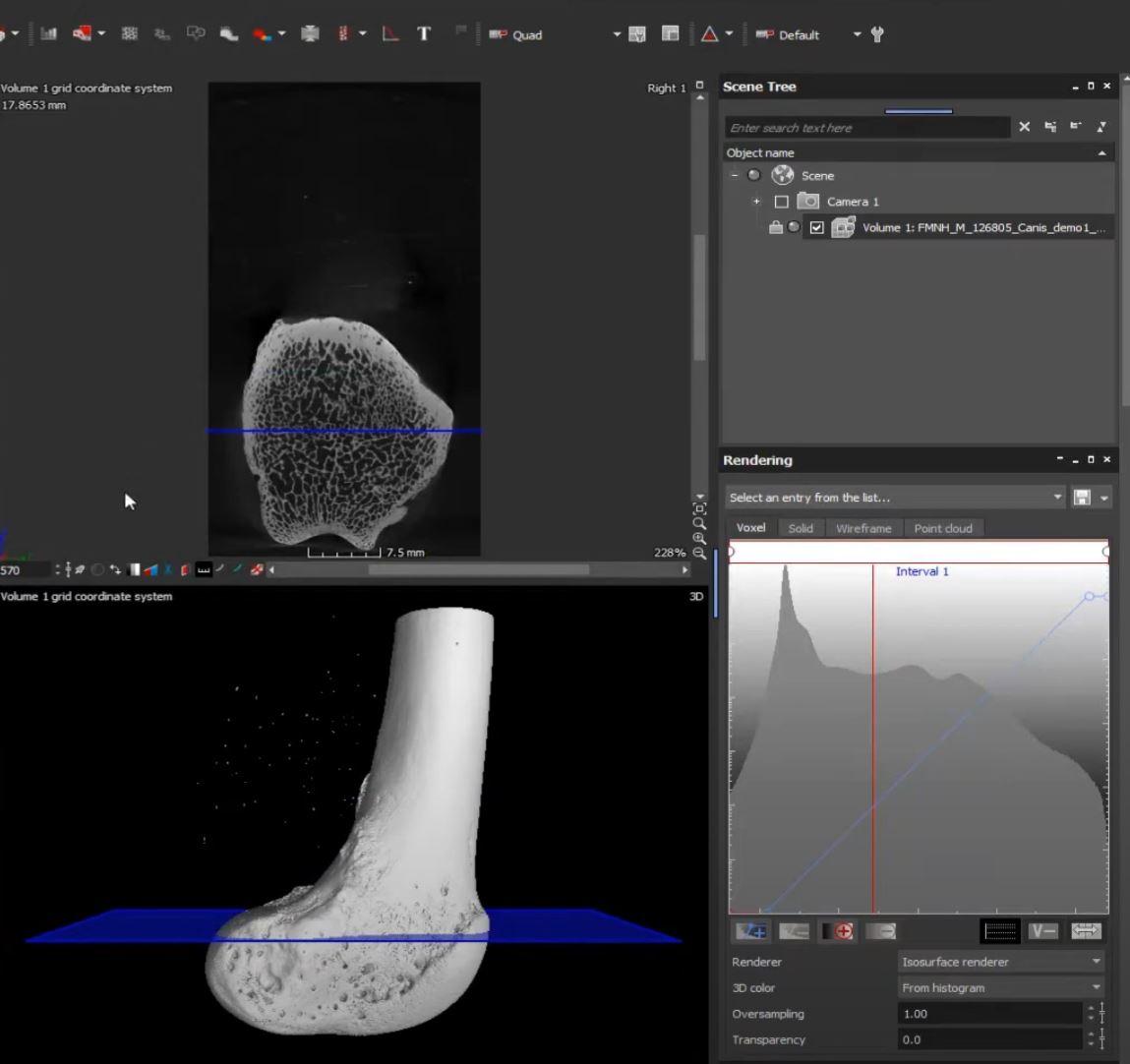
By April Neander, The University of Chicago Chapter 12 of the University of Chicago PaleoCT training course. Covers importing data, navigation, reorientation, cropping, and merging scans in VGStudio. https://youtu.be/oWecQ6AmN2A
Educational Resource
Posted on:
Basics and Physics of X-Ray Imaging & 3D Computed Tomography (Volume Graphics)
By S. Gondrom-Linke, Volume Graphics https://www.dropbox.com/s/zlvqvtklmqdu3d3/Physics%20and%203DCT.pdf?dl=0 CT history, basics, how a CT scanning and reconstruction works with relevant mathematical explanations, and demonstration of how VGStudio Reconstruction can be used to correct artifacts.
Educational Resource
Posted on:
NC State University Analytical Instrument Facility: Short Introduction to X-ray Computed Tomography
Video about basics of how CT works. https://youtu.be/liVAgLJmq8g
Educational Resource
Posted on:
X-Ray Microscopy Introduction
By Jon Giencke, Bruker X-ray Diffraction (XRD) and X-ray Microscopy (XRM) can provide a wealth of information while being non-destructive and easily accessible. This video series includes an exciting line-up of speakers to discuss the basics of the techniques, provide […]
Educational Resource
Posted on:
Micro-computed tomography for natural history specimens: a handbook of best practice protocols
Keklikoglou, Kleoniki, Sarah Faulwetter, Eva Chatzinikolaou, Patricia Wils, Jonathan Brecko, Jiří Kvaček, Brian Metscher, and Christos Arvanitidis. “Micro-computed tomography for natural history specimens: a handbook of best practice protocols.” European Journal of Taxonomy 522 (2019). https://doi.org/10.5852/ejt.2019.522 Abstract Micro-computed tomography (micro-CT or microtomography) […]
Educational Resource
Posted on:
Artefacts in X-ray microtomography of materials
Davis, G. R., and J. C. Elliott. “Artefacts in X-ray microtomography of materials.” Materials science and technology 22, no. 9 (2006): 1011-1018. https://doi.org/10.1179/174328406X114117 Abstract X-ray microtomography is becoming an increasingly popular tool in the study of microstructure and failure mechanisms in biological […]
Educational Resource
Posted on:
Seattle Children’s Research Institute
By Murat Maga, Seattle Children’s Research Institute https://faculty.washington.edu/maga/SCRI_MicroCT/ Facility Website that offers CT scanning services, hosts mouse CT Data for download, 3D Visualizations, and other useful documents.
Educational Resource
Posted on:
Spatial Archaeometry Research Collaborations (SPARC) Teaching Resources for microCT (University of Arkansas)
By Manon Wilson, University of Arkansas https://github.com/castuofa/sparc_microct Educational resources including CT Data analysis and visualization focused on the field of archaeology.
Educational Resource
Posted on:
Enhancing CT imaging: A safe protocol to stain and de-stain rare fetal museum specimens using diffusible iodine-based staining (diceCT)
Lanzetti, Agnese, and Eric G. Ekdale. “Enhancing CT imaging: A safe protocol to stain and de‐stain rare fetal museum specimens using diffusible iodine‐based staining (diceCT).” Journal of Anatomy 239, no. 1 (2021): 228-241. https://doi.org/10.1111/joa.13410 Abstract Computed tomography (CT) scanning is being increasingly […]