
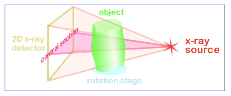
MicroCT imaging, short for micro-computed tomography, is a technique that uses X-rays to create detailed 3D images of objects. It works by taking many 2D X-ray images, called projection images, of an object from different angles and then combining them using a process called reconstruction to form a 3D virtual histology of the object. This method is non-destructive, meaning the object being scanned remains intact.
Thank you to Murat Maga at the Seattle Children’s Research Institute’s Center for Developmental Biology and Regenerative Medicine for organizing this tutorial.